CONTROL STRATEGIES OF SHIFTING
DURING VIOLIN PLAYING
by
Lauren Michelle Deutsch
A Thesis Presented to the
FACULTY OF THE
In Partial Fulfillment of the
Requirements for the Degree
MASTER OF SCIENCE
(KINESIOLOGY)
May 2006
Copyright 2006
Lauren
Michelle Deutsch
TABLE OF CONTENTS
| List of Tables and Figures | |
| Abstract | |
| Chapter 1: Introduction | |
| Chapter 2: Methods | |
| Subject Selection | |
| Task Description | |
| Experimental Protocol | |
| Data Processing and Analysis | |
| Chapter 3: Results | |
| Comparisons Within Subject, between Tasks | |
| Subject 1: EMG Data | |
| Subject 1: Kinematics Data | |
| Subject 2: EMG Data | |
| Subject 2: Kinematics Data | |
| Subject 3: EMG Data | |
| Subject 3: Kinematics Data | |
| Comparisons Between Subjects, Within Tasks | |
| Physical Evaluation Results | |
| Chapter 4: Discussion | |
| Bibliography |
ABSTRACT
Overuse injuries are common among violinists. Due to the awkward postural position required, violinists often suffer from injuries to the upper extremities. The aim of this research was to determine how experienced violinists control the left scapula and upper extremity during playing tasks performed along the length of the fingerboard using their preferred technique. Activation of muscles controlling the scapula and left upper extremity was monitored (surface electromyography, 2400 Hz) and segment kinematics were recorded (digital video, 60 Hz). Comparison of muscle activation patterns within each subject across tasks revealed that individual players scale different sets of muscles to accommodate playing in different positions along the fingerboard. Comparison of muscle activation patterns between subjects (n=3) within tasks revealed that two different neuromuscular control strategies were used to control scapular and upper extremity motion during the same playing tasks.
Chapter 1: Introduction
Overuse injuries
are common among violinists. According to a survey conducted by the
International Conference of Symphony and Opera Musicians in 1986, eighty-two
percent of musicians reported experiencing a medical problem and seventy-six
percent of those surveyed reported a problem that required time off from
playing 5. Due to the awkward postural position required for playing
the violin, violinists often suffer from injuries to the upper extremities, and
violinists as a group suffer more injuries to their left arm than their right 10.
Common left upper extremity injuries include rotator cuff tears, tendonitis,
thoracic outlet syndrome, carpal tunnel syndrome, and nerve entrapments 5.
Presently, there is not very much scientific data available regarding the mechanics and the neuromuscular control of the left upper extremity during violin playing. The majority of the published information on the subject is in the form of treatises by pedagogues who give suggestions on violin technique based on years of teaching experience 2,3,7,22. There are a handful of studies that collected muscle activation patterns during violin playing 9,12,17,18; however, none of the studies have combined the use of EMG and kinematics to study control strategies used by violinists. In addition, many past experimental studies have focused on investigating the prevalence or reporting on the treatment of injuries commonly suffered by musicians. One such study reported the use of physical therapy and medicine to help treat musicians after the onset of an injury 10. For example, thoracic outlet syndrome patients are treated by initially modifying harmful playing positions through facilitation of instrument adaptations (i.e. shoulder rests, chin rests, etc.) and then given physical therapy to strengthen and stretch the affected areas 10. In addition, there have been books written with suggestions on injury prevention for violinists. In Janet Horvath’s book, Playing (less) Hurt, she recommends that violinists experiment with shoulder pads and chin rests to achieve a position that does not require raising the shoulder up or thrusting it forward 5.
Analysis of segment kinematics and muscle activation patterns during the performance of goal directed tasks has provided valuable insight as to load distribution during well-practiced goal-directed tasks. Muscle activation patterns provide evidence regarding strategies the neuromuscular system uses to control posture as well as segment motion 1. It is expected that monitoring upper extremity kinematics and muscle activation patterns during violin playing will advance our understanding of how left arm movement is coordinated and controlled 23.
Since the most fundamental left hand technique that is required of violinists as they advance in skill is shifting (movement of the left hand along the neck of the violin to reach high and low notes), playing tasks in positions along the length of the fingerboard will be chosen for study. By comparing muscle activation of scapular and upper extremity muscles (using surface electromyography) and kinematics of the scapula and upper extremity (using 60 Hz video), we will determine the neuromuscular control and joint motion used by advanced violinists to play in different positions along the neck of the violin. Motion of the left upper extremity during shifting is constrained by the fact that the fingers must contact the strings along the fingerboard. As a result, the violin player utilizes available degrees of freedom afforded by the scapula and upper extremity to achieve enough rotation of the arm and hand so that the fingers can reach and depress the strings along the entire length of the fingerboard. Playing on the lowest string (the G string) presents the greatest challenge to the neuromuscular system of the upper extremity by requiring the most rotation in order to reach and depress the string.
The aim of this research was to determine how experienced violinists control the left scapula and upper extremity during playing tasks performed along the length of the fingerboard using their preferred technique. It was hypothesized that: a) playing tasks performed along the length of the fingerboard would require different neuromuscular control patterns and b) different violin players would exhibit unique muscular control patterns when performing the same playing tasks. It was expected that violinists adopt control strategies that fit within their physical capabilities and preferred playing position; consequently, it is probable that subjects will engage in unique control strategies. Since external rotation of the shoulder, supination of the forearm, and/or tilt of the scapula are the degrees of freedom available to the subjects to accomplish the tasks (play on the G string in the 1st, 3rd, and 10th positions), subjects with limitations in the range of movement of these joints must compensate by finding an alternate control strategy.
All violin tasks involved depression of the G string along the fingerboard of the violin (specifically 1st, 3rd, and 10th positions; Figure 1). These tasks were selected in that they were expected to impose the greatest demands on the neuromuscular system. Muscle activation patterns of muscles controlling the scapula and left upper extremity were monitored using surface electromyography. Segment kinematics during violin playing were analyzed using digital video. Results of this research provide evidence for identifying techniques for effectively distributing mechanical load during violin playing. The basic foundation of knowledge established in this study will be necessary for designing effective future studies aimed at gaining a more in-depth understanding of the possible causes and treatments that can be put into practice by injured violinists.
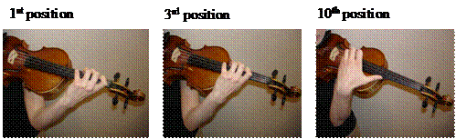
Figure 1: 1st, 3rd, and 10th positions
CHAPTER 2: METHODS
Subject
Selection
Three
female violinists from the Los Angeles
Task
Description
Each subject was asked to play a simple pattern of eighth-notes to a metronome set at 60 beats per minute in 1st, 3rd, and 10th positions on the G string (Figure 1). The fingers were played in the sequence 1st, 2nd, 3rd, 3rd, with whole steps in between each note. Therefore, the task in 1st position included the notes A, B, C#, C#; the task in 3rd position included the notes C, D, E, E; the task in 10th position included the notes C, D, E, E (one octave above the third position pitches). Each task was performed three times each. The subjects performed the trials in a seated position and were instructed to hold the violin in their “preferred” manner (they used their own instrument and accessories, and they could hold their violin at any angle or height they chose to).
Experimental
Protocol
Activity of
representative uni- and bi- articular left upper extremity muscles were monitored
using telemetered surface electromyography (EMG) (2400 Hz; Beckman
Silver-Silver Chloride electrodes; Konnigsberg,
Three-dimensional kinematics
was recorded using five 60 Hz video cameras (60 fps; Sony). Reflective markers
were placed on the scapula (Trigonum Spinae, Inferior Angle, and Angulus
Acromialis), lower arm (medial epicondyle, lateral epicondyle, ulnar styloid
process, radial styloid process), upper arm (3 non-colinear points were
attached to a rigid cuff), and hand (below the 5th metatarsal, below
the 2nd metatarsal, on the rigid hand segment) to facilitate the
process of kinematic analysis 21. Cameras were placed to the
front-right, front-left, back-right, back-left, and overhead of the subject.
The video analysis software (TeamPro 4.0.3.0, Dartfish) was used to overlay
videos of the different tasks to make qualitative comparisons as well as to
measure the preferred violin angle of the different subjects (using the
overhead video clips).
Subjects also underwent a physical evaluation to determine the range of motion of the forearm and shoulder (i.e. internal rotation, external rotation, and supination). Standard physical therapy assessment techniques were used for these evaluations 7.
Data
Processing and Analysis
All EMG data was band-passed filtered (10-400 Hz) using a fourth order, recursive Butterworth filter. The root-mean-square (RMS) of the EMG data was binned in 10ms intervals. After normalizing the data to the maximum (filtered and binned) maximum muscle activation during the manual muscle test of each muscle, averages were taken for each muscle over the three seconds of the task, establishing a normalized average RMS value representative of the amount of activity of the muscle during that particular trial. The above process was repeated for all three trials in all three positions for each subject. After getting an average RMS value for all three trials in one position, these values were averaged to represent the average amount of activity of a particular muscle in a particular position. This resulting number was used to express the amount of normalized EMG activity in each muscle expressed as a percent of the MMT task. Standard deviations of the three trial average RMS values were also calculated for each muscle. The data was analyzed in two ways: 1) The amount of muscle activity in positions 1, 3, and 10 (for each muscle) was compared within each subject to determine whether there were “between-task, within subject” differences in neuromuscular control patterns. 2) Muscle activation patterns for each subject were compared to determine if there were “between-subject, within task” differences in the neuromuscular control patterns and whether these differences can be explained through the findings in the kinematics data and physical evaluation.
CHAPTER 3: RESULTS
Comparisons
Within Subject, Between Tasks
Comparison of muscle activation patterns within subject across tasks revealed that individual players scale different sets of muscles when depressing the G-string in 1st, 3rd, and 10th positions.
Subject
1: EMG Data
Subject 1 showed similar “between-task” muscle activation patterns for all muscles except the UT and the FF (Figure 2). Muscles that showed similar “between-task” activation patterns (differences between tasks did not exceed beyond the range of the standard deviation between trials) included the P Major, Lat, A Delt, PD, MD, Inf, SA, MT, LT, SBc, LBc, LTr, LatTr, and FE. Muscles that exhibited activation levels of greater than 20% included the P Major, MD, Inf, MT, and SBc.
The average UT activity exhibited the tendency to decrease as the hand moved closer to the body along the fingerboard (from 1st position to 10th position). The UT activity was highest (8.2 ± 0.03%) when playing in 1st position, an average of 17.7% lower (6.8 ± 0.72%) than when playing in 3rd position, and lowest (4.5 ± 0.66%) when playing in 10th position. The average FF activity was similar when playing in 1st and 3rd positions (9.0 ± 0.03% and 9.5 ± 1.09% respectively); however, the average FF activity when playing in 10th position (11.9 ± 0.31%) was 24.73% greater than in 3rd position.
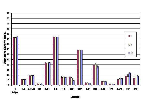
Figure 2: Subject 1’s EMG activity in 1st, 3rd, and 10th positions
Subject
1: Kinematics Data
Subject 1’s segment kinematics in 1st position included abduction, external rotation, and flexion of the shoulder, flexion of the elbow, and supination of the forearm (Figure 3a). The kinematics in 3rd position are similar to the kinematics in 1st position with the exception that the elbow is flexed to a greater degree (Figure 3b). The kinematics in 10th position are similar to the kinematics in the 1st and 10th positions; however, flexion of the wrist is now present, and there is a greater degree of elbow flexion, shoulder flexion, shoulder external rotation, and forearm supination (Figure 3c).
Figure 3: Subject 1’s Kinematics Data in 1st, 3rd,
and 10th positions
Subject
2: EMG Data
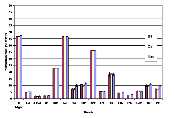
Figure 4: Subject 2’s EMG activity in 1st, 3rd,
and 10th positions
Subject 2 showed similar “between-task” muscle activation patterns for all muscles except the SA (Figure 2). Muscles that showed similar “between-task” activation patterns (differences between tasks did not exceed beyond the range of the standard deviation between trials) included the P Major, Lat, A Delt, MD, Inf, UT, MT, LT, SBc, LBc, LTr, LatTr, FF, FE (Figure 4). Muscles that exhibited activation levels of greater than 20% included the P Major, MD, Inf, and MT. Activity of the SA increased by 32.57% in the 10th position (9.85 ± 0.84%) as compared to the similar activation levels of the SA in 1st (7.31 ± 0.30%) and 3rd position (7.43 ± 0.75%).
Subject
2: Kinematics Data
Subject 2’s segment kinematics in 1st position included abduction, external rotation, and flexion of the shoulder, flexion of the elbow, and supination of the forearm (Figure 5a). The kinematics in 3rd position are similar to the kinematics in 1st position with the exception that the elbow is flexed to a greater degree (Figure 5b). The kinematics in 10th position are similar to the kinematics in the 1st and 10th positions; however, flexion of the wrist is now present, and there is a greater degree of elbow flexion, shoulder flexion, and shoulder external rotation (Figure 5c).
 Figure 5:
Subject 2’s Kinematics data in the 1st, 3rd and 10th
positions
Figure 5:
Subject 2’s Kinematics data in the 1st, 3rd and 10th
positions
Subject
3: EMG Data
Subject 3’s EMG
exhibited large standard deviations when averaging all three trials together
especially when playing in 3rd position (Figure 6). For example, the standard deviation of the
three trials for the LBc data in 3rd position
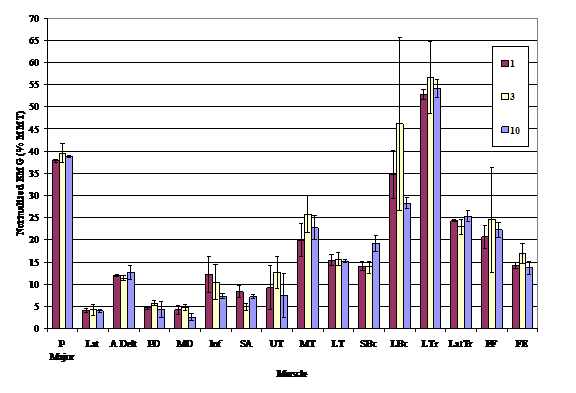
Figure 6: Subject 3’s EMG activity in 1st, 3rd, and 10th positions (all trials included)
was 19.56%. This high standard deviation in the LBc in 3rd position was also accompanied by a high standard deviation in the LTr (8.09%) and FF (11.83%) in 3rd position.
Trial 3 of the 3rd position task contributed to this high standard deviation (Figure 7). These results indicate that the third trial performed in 3rd position was not representative of exemplar task performance (Figure 7). Therefore, only the first two trials were used for analysis of trends in the average EMG data for this subject. After the removal of the third trial performed in 3rd position, the standard deviations decreased to 1.56% in the LBc, 1.0% for the LTr, and .02% for the FF (Figure 8).
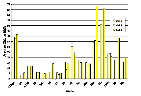
Figure 7: Subject 3’s EMG activity in all 3rd position trials
Using average EMG data including only trials 1 and 2 (Figure 8), slight between-task differences were observed in the SA and FF whereas larger between-task differences were observed in the SBc and LBc. The SA activity was lowest in 3rd position (4.78 ± 1.04%), higher in 10th position (7.18 ± 0.42%), and highest in 1st position (8.35 ± 1.38%). The FF activity was lowest in the 3rd position (17.73 ± 0.02), higher in 1st position (20.66 ± 2.52%), and highest in 10th position (22.30 ± 1.68%). The SBc activity was similar in 1st and 3rd positions (approximately 14%) and higher in 10th position (19.21 ± 1.86%). Similarly, the
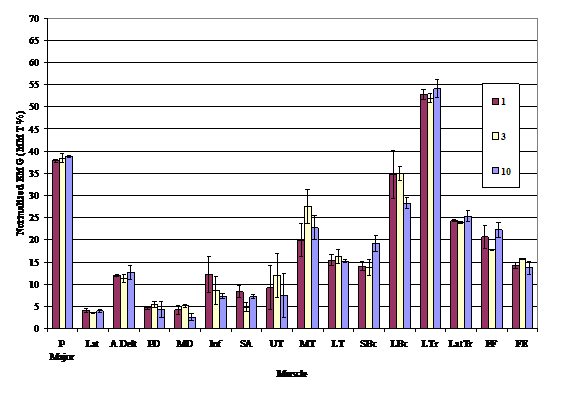
Figure 8: Subject 3’s EMG activity in 1st, 3rd, and 10th Positions (after removal of the 3rd trial of the 3rd position task.)
LBc activity was similar in 1st
and 3rd positions (approximately 35%); however the LBc activity
decreased in 10th position to 28.22 ± 1.24%.
Subject 3: Kinematics
Data
Subject 3’s segment kinematics in 1st position included abduction, external rotation, and flexion of the shoulder, flexion of the elbow, and supination of the forearm (Figure 9a). The kinematics in 3rd position are similar to the kinematics in 1st position with the exception that the elbow is flexed to a greater degree (Figure 9b). The kinematics in 10th position are similar to the kinematics in the 1st and 10th positions; however, flexion of the wrist is now present, and there is a greater degree of elbow flexion, shoulder flexion, and shoulder external rotation (Figure 9c). All 3 trials in the 3rd position exhibited similar kinematics.
 Figure 9:
Subject 3’s Kinematics data in the 1st, 3rd and 10th
positions.
Figure 9:
Subject 3’s Kinematics data in the 1st, 3rd and 10th
positions.
 Figure 10: 3rd
Position Individual Trial Kinematics
Figure 10: 3rd
Position Individual Trial Kinematics
Comparisons
Between Subjects, Within Tasks
Comparison of the “between-subject” activation patterns support the original hypothesis that different violin players would exhibit unique muscular control patterns when performing the same playing tasks. While both subject 1 and 2 utilized similar muscular control patterns, subject 3 utilized a different muscular control pattern (compared to subjects 1 and 2) when performing the playing tasks.
Both subjects 1 and 2 utilized relatively high activation levels of the muscles controlling the shoulder (MT) and shoulder girdle (MD and Inf), while utilizing relatively low levels of the muscles controlling the shoulder and elbow (LBc, LTr, and LatTr) during the playing tasks. In contrast, subject 3 utilized relatively high activation levels of the muscles controlling the shoulder and elbow (LBc, LTr, and LatTr), while utilizing relatively low activation levels of the muscles controlling the shoulder girdle and shoulder (Inf, MT, and MD). However, all subjects utilized relatively high levels of the P Major (controlling the motions of flexion, internal rotation, and adduction of the shoulder joint) and the SBc (controlling the motions flexion and adduction of the shoulder as well as flexion of the elbow and supination of the forearm).
Physical
Evaluation Results
|
Subject |
Supination (°) |
External Rotation (°) |
Internal Rotation (°) |
Violin Angle Preferred (°) |
|
1 |
103.9 |
76.0 |
57.3 |
57.4 |
|
2 |
90.4 |
60.2 |
36.0 |
67.5 |
|
3 |
118.9 |
58.9 |
32.4 |
59.0 |
Table 1:
CHAPTER 4: DISCUSSION
The purpose of this study was to advance understanding of the neuromuscular control of the left upper extremity during violin playing. To determine how experienced violinists control the left scapula and upper extremity during violin playing tasks, the muscle activation patterns and segment motion were monitored and compared between playing tasks performed along the length of the fingerboard. It was hypothesized that: a) playing tasks performed along the length of the fingerboard would require different neuromuscular control patterns and b) different violin players would exhibit unique muscular control patterns when performing the same playing tasks. By monitoring upper extremity kinematics and muscle activation patterns during violin playing in 1st, 3rd and 10th positions along the fingerboard, between-task differences and between-subject differences in control were observed. Comparison of muscle activation patterns within subject across tasks revealed that individual players scale different sets of muscles when depressing the G-string in 1st, 3rd, and 10th positions. Comparison of muscle activation patterns between subjects, within tasks revealed that two different neuromuscular control strategies were used to control scapula and upper extremity motion during playing tasks. Two players tended to utilize control strategies that disproportionately activated muscles controlling the shoulder and shoulder girdle, whereas a third violin player used a strategy that disproportionately activated muscles controlling upper extremity during the same playing tasks.
Even with the limitations of a small sample size (n=3), the experimental results demonstrate that there is more than one way to control playing tasks in different positions along the length of the fingerboard. While all subjects had been playing the violin for over 10 years, there were differences in experience, practice schedules, and physical characteristics. For example, subject 1 had been playing almost twice as long as subject 3 partially because of the 15 year difference in chronological age. In addition, the subjects that participated in the study were at different stages in their career. Subject 3 was still a student taking regular weekly lessons and practicing approximately 3 hours per day, subject 1 was practicing and/or playing between 1 and 3 hours per day at the time of the study, and subject 2 was not on a regular practice schedule during the months leading up to the study. Subject 3 reported a history of pain in the shoulder/scapular region, whereas subjects 1 and 2 did not report any history of pain. Because this study focused on the muscle activation patterns individuals use during everyday violin playing, each subject was entitled to select their own playing position and violin position. As a result, the effect of violin tilt, violin angle, chinrest position, and shoulder pad characteristics were allowed to vary according the preferences of the individual violin players. While future experiments can be designed to determine the effect of these characteristics on muscle activation patterns, the experimental data acquired in this study reflects the muscle activation used by the individual under their preferred playing conditions. As such, the results of this study reflect neuromuscular control patterns used during violin playing under typical practice conditions.
Within subject, between-task differences in muscle activation patterns were consistent with how the individual player chose to shift from position to position. For example, subject 1 showed a slight trend of decreased UT activity as the fingers moved away from the body. The highest amount of UT activity was observed in 1st position and the lowest amount of UT activity was observed in 10th position. One possible reason for this decreasing trend in UT activation could be that the subject elevated or upwardly rotated her scapula more in the 1st position as compared to the 10th position. Since the scapula kinematics are essentially the same between 1st and 10th positions, differences in scapular positions as the source of this observed difference in UT activation levels is discounted. While the relationship between UT activation and the playing tasks suggests that subject 1 uses the UT in the regulation of playing along the fingerboard, the magnitude of the muscle activation is minimal in comparison to the activity observed during the MMT task. Therefore, the change in kinematics may not be detectable using simple video overlays, since scapular changes are difficult to measure using tracking markers attached to the skin because of motions that occur underneath the skin 21.
Similarly, subject 2 exhibited increased SA activity in 10th position (compared to 1st and 3rd positions). Since the SA is responsible for abducting and upwardly rotating the scapula, one would expect the scapular kinematics to show more abduction and upward rotation of the scapula. However, just as in subject 1, the scapular kinematics in subject 2 do not show any changes from 1st and/or 3rd positions compared to the 10th position. In addition, the magnitude of muscle activation of the SA during the playing tasks is minimal in comparison to the activity observed during the MMT task; therefore, using the same reasoning as subject 1, the scapular kinematic changes may not have been observable using video overlays.
Due to the construction of the violin, between-task differences in muscle activation levels were also attributed to the relative difficulty of depressing the G string down to the fingerboard in 10th position relative to the 1st and 3rd positions. For example, subject 1 exhibited an increase in the FF activation in 10th positions as compared to the 1st and 3rd positions reflecting the increase in distance from the string to the fingerboard as the position increases.
Subject 3 showed the most variability in EMG activity between tasks and trials. The source of between-trial variability in activation levels of the FF, LBc, and LatTr is consistent with regulating force associated with string depression. Variability in the FF was most pronounced in the 3rd position trials (FF activation was more than two times greater in trial 3 than in trials 1 and 2). Since the increase in FF activation in trial 3 was accompanied by a simultaneous increase in LBc and LatTr activity, perhaps these muscles (FF, LBc, and LatTr) are associated with force regulation (how hard to press the string down) in this subject. However, since force data was not collected, this explanation cannot be verified directly.
The results of this study indicate that more than one neuromuscular control strategy can be used by violinists to perform playing tasks. Two of the three violin players utilized a shoulder and shoulder girdle control strategy, while one subject utilized an arm control strategy. Subjects 1 and 2 both used control strategies that involved activation of mainly the shoulder girdle (MT) and shoulder muscles (Inf and MD), while Subject 3 utilized a control strategy that involved activation of mainly the arm muscles (LBc, LatTr, and LTr). The emergence of the two unique muscle activation patterns was unaccompanied by any obvious differences in the kinematics between subjects 1, 2, and 3; therefore, subjects accomplished similar segment motions through the activation of different sets of muscles.
While these two different control strategies may signify that emergence of two possible control strategies that can be used to play the violin along the length of the fingerboard, more subjects would be needed to rule out other possible reasons for the emergence of these two different control strategies. For example, subject 3 reported a history of pain in the shoulder and scapular region. Therefore, subject 3 may have chosen to control playing tasks with the arm rather than scapular muscles in order to avoid previously occurring pain in the shoulder and shoulder girdle areas.
One possible explanation for the two control strategies involves using different sets of muscles to position the humeral head in the glenoid fossa to provide stabilization of the shoulder joint. While subjects 1 and 2 may have utilized high activation levels of the Inf to increase the stability and decrease superior translation of the glenohumeral (shoulder) joint in the glenoid fossa, subject 3 may have substituted use of the Inf with use of the LBc, which also can function in the same way as the Inf to stabilize the glenohumeral joint 11. However, since the LBc also performs other functions (such as abduction and flexion of the shoulder, flexion of the elbow, and supination of the forearm), the LatTr (extension of the elbow) and LTr (extension and adduction of the shoulder as well as extension of the elbow) accompanied activation of the LBc to counteract all of the segment motions that were unnecessary for the playing task. Since the triceps muscle group (LTr and LatTr) opposed additional actions of the LBc that were not necessary for the playing tasks, the shoulder and elbow positions were stable when fingering in each position. This logic can also explain why the LBc showed decreased activation in 10th position, while the SBc showed increased activation in 10th position – since the arm was closer to the body in 10th position, the humeral head may not have needed as much force from the LBc for stabilization and instead the SBc activated at a higher level to provide more elbow flexion for the 10th position.
Another possible explanation for the difference in control strategies include links between the physical characteristics and the control strategy chosen; however, this study does not show any clear correlations between physical characteristics of the subjects and the control strategy utilized. While subjects 2 and 3 had similar physical plant limitations in external and internal rotation of the shoulder, they did not choose the same control strategy. Consequently, the physical evaluation data is insufficient for determining muscle recruitment patterns in these subjects.
While the specific benefits and limitations of the arm control strategy versus the shoulder control strategy are unknown, time to muscle fatigue has been found to occur more quickly when the activation is a higher percentage of the maximum voluntary contraction (MVC) of that muscle 15. Monod found that the “critical force,” the point where the time to fatigue increases exponentially, lies around 20% of MVC. Since risk of overuse injury is thought to increase when a muscle is fatigued 5, muscle activation levels over this threshold should be noted. Therefore, subjects 1 and 2 may be susceptible to overuse of the P Major, Inf, MD, and MT, whereas subject 3 may be susceptible to overuse of the LBc, LTr, LatTr, P Major, and MT. While future studies would be needed to confirm this link, future treatments for overuse injuries could train subjects to combine control strategies so that all muscles are being used at a lower activation level so that none of the muscles would be taxed disproportionately. In addition, this information would encourage violinists to participate in muscle strengthening activities so that their maximum voluntary contractions of the muscles used during violin playing would increase, resulting in decreased percentages of the maximum that would be needed during violin playing.
This study raises many questions. For example, if more violinists were studied, would we find an equal distribution among violinists who use the shoulder and shoulder girdle versus the arm control strategy? Would we discover additional strategies that are used by violinists? Future research can address some of these questions to clarify the findings of this study. All information increasing the understanding of how the left upper extremity is controlled during violin playing will help to find ways of treating and preventing the high incidence of left upper extremity injuries in violinists.
BIBLIOGRAPHY
1. De Luca, Carlo J. “The Use of Surface Electromyography in Biomechanics.” Journal of Applied Biomechanics 13 (1997): 435-465.
2. Flesch,
Carl. The Art of Violin Playing.
Trans. and ed. Eric Rosenblith, with a foreword by Anne-Sophie Mutter.
Book 1.
3. Galamian,
Ivan. Principles of Violin Playing and
Teaching. 3d ed.
4. Hodgson, Percival. Motion Study and Violin Bowing. n.p. American String Teachers Association, 1958.
5. Horvath,
Janet. Playing (less) Hurt: An Injury
Prevention Guide for Musicians. 3d ed.
6. Jackson, K. M. “Fitting of Mathematical Functions to Biomechanical Data.” IEEE Trans Biomed Eng 26 (1979), 122-124.
7. Kempter,
Susan. How Muscles Learn: Teaching the
Violin with the Body in Mind.
8.
9.
Kjelland,
James M. “Application of Electromyography and
10. Lederman,
R.J. “Neuromuscular and Musculoskeletal Problems in
11. Levangie,
Pamela K. and Cynthia C. Norkin. Joint
Structure and Function: A
12. Levy, Charles E., W.A. Lee, A.G. Brandfonbrener,
J. Press, and A.E. Levy.
13. Ludewig,
Paula & Thomas Cook. “Alterations in Shoulder Kinematics and
14. Medoff, Lynn. “The Importance of Movement Education in the Training of Young Violinists.” Medical Problems of Performing Artists 14 (1999), 210-218.
15. Monod, Hugues. “Contractility of Muscle during Prolonged Static and Repetitive Dynamic Activity.” Ergonomics 28, no. 1 (1985), 81-89.
16. Mozart,
Leopold. A Treatise on the Fundamental
Principles of Violin Playing Trans. Editha Knocker. 2nd ed.
17. Philipson, L., R. Sorbye, P. Larsson, and S. Kaladjev. “Muscular Load Levels in Performing Musicians as Monitored by Quantitative Electromyography.” Medical Problems of Performing Artists 5 (June 1990), 79-82.
18. Polnauer,
Frederick and Marks, Morton. Senso-motor
Study and It's Application
to Violin Playing.
19. Szende,
Otto, and Mihaly Nemessuri. The
Physiology of Violin Playing Trans. I. Szmodis, with a foreword
by Yehudi Menuhin and preface by Paul Rolland.
20.
21. Van der Helm, Frans C.T. 1997. A standardized protocol for motion recordings of the shoulder. Proceedings of the First Conference of the International Shoulder Group.
22. Vigdorchik,
Isaak A. Violin Playing: A Physiological
Approach.
23. Zatsiorsky, Vladimir. Kinematics of Human Motion. n.p. Human Kinetics, 2002.